


Synapses spotted on the move
 Print
Print
 Australian researchers have caught the first ever glimpse of the brain’s tiniest molecules in action.
Neuroscience researchers at the University of Queensland used a breakthrough technique in super-resolution microscopy to spot and plot the movements of molecules within synapses.
The technique was developed at Professor Fred Meunier’s laboratory at UQ's Queensland Brain Institute.
“The technique will be revolutionary for neuroscientists and cell biologists,” Professor Meunier said.
“This is an exciting time, as we’ve opened the door for many more ground-breaking studies that will change our view of how molecules function to make the brain work.
“These images could further our understanding of memory, learning, and neurodegenerative diseases.”
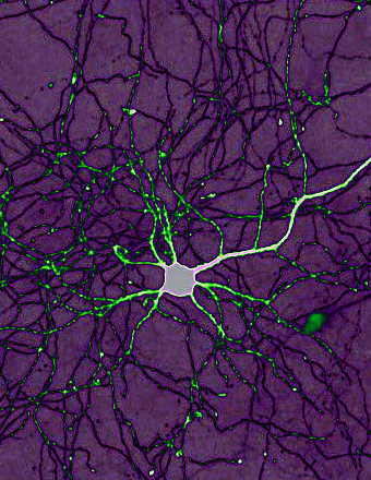
The breakthrough imaging technique captures the movement of molecules within synapses – structures connecting brain cells – that are too small for conventional microscopes.
Classical optical microscopes can only clearly identify objects that are 200 nanometres (1/5000th of a millimetre).
Super-resolution microscopy techniques can achieve 10 to 20 times higher resolution (closer to electron microscopy), even on living cells.
“You see molecules moving randomly and then, for a few milliseconds, interacting with each other.
“Control of these critical moments define neurotransmission and allow brain cells to communicate efficiently.”
The technique has led to a series of papers from Professor Meunier’s team published in the Journal of Cell Biology and Nature Communications, including;
- Tracking single molecules in living brain cells undergoing communication
- Tracking these molecules within brain cells of living organisms such as fruit flies
- Tracking single tiny structures, called synaptic vesicles, that are responsible for the release of neurotransmitters at the synapse
- Imaging tiny signalling endosomes responsible for brain cell survival as they move from the synapse to the cell body.